

Computer-Aided Diagnosis In Dermatology
Computer-Aided Diagnosis for Skin Cancer: Skin cancer is one of the common cancers in the world; if detected early, it can be treated. Dermatology is primarily dependent on visual assessment skills, especially with increased patient load and limited professional availability; there is a need for greater accuracy in detection methods.
The traditional methods were supplemented by smarter digital tools, and the development of computer-aided diagnosis is now transforming the field, helping clinical experts to identify suspicious lesions early.
Dermatology has become one of the most active areas of technological advancements with the rise of artificial intelligence in healthcare. A combination of various diagnostic tools, like AI skin analysis, medical image analysis, and dermatology imaging technology, is now transforming everything from smartphone-based triage to advanced dermoscopic evaluation.
The Need for Better Skin Cancer Detection Tools:
Early detection of skin cancer helps in assessing lesions; moreover, many benign lesions mimic malignant ones, posing diagnostic challenges for clinicians and pathologists because they appear the same on physical examination. Along with these high patient volumes, short consultation times, there is limited access to dermatologists in rural areas, which is another challenge.
This is where AI and other advanced imaging tools come into the picture, making a measurable difference. Using artificial intelligence and machine learning in dermatology helps in automation, like the detection of skin lesions. These advancements help reduce the burden on clinicians.
AI helps to shift from reactive to proactive care when it is combined with predictive analysis in healthcare. This helps clinical professionals to create strategies to identify which lesions and which patients may require what type of examination frequently.
How computer-aided diagnostics works in dermatology:
Computer-aided diagnostics (CAD) in dermatology is primarily designed to support clinicians in analysing the images, identifying the patterns, and assessing their risk levels. The workflow combines several interconnected technologies:
- Image Capture and Standardization:
A dermatologist uses dermascopes, smartphone attachments, and high-resolution clinical imaging systems for high-quality imaging. This helps in constant lighting and focus to ensure reliable medical imaging.
- Preprocessing and Artifact Correction:
In this step, the lesion remains in the focal point. Before the analysis, AI systems make sure that they clean the image by removing hair, noise, shadows, and reflections for accurate and precise treatment.
- Feature Extraction:
This step involves the use of deep learning for medical imaging. It uses AI models/systems to identify and detect the colour differences, patterns in the pigment, distortions in the structure, vascular clues, and border irregularities.
- Classification and Scoring:
Here, AI tools are used to categorize lesions into different categories, like low, moderate, and high risk. This does not replace clinicians but helps to improve their choice of making decisions. This is where clinical decision support systems become valuable, providing information during examination.
Computer-aided diagnostic systems improve overall accuracy, consistency, and efficiency. Especially for reducing the burden on clinicians and for regions with limited dermatology resources.
AI Dermatology Tools for Smartphone-Based Screening:
Integration of AI dermatology tools into smartphones is one of the transformative advancements, where modern mobile cameras, that are supported by cloud-based AI tools, help in the assessment outside traditional hospitals.
Benefits of Smartphone AI in Dermatology:
- People in the remote areas can easily access and screen the suspicious lesions.
- Physicians/clinicians can easily identify the cases that need urgent attention/emergency treatment.
- People can also track the changes in the lesions over time.
- It can be used for educational purposes, as students and trainees can learn visual patterns through the guided tools.
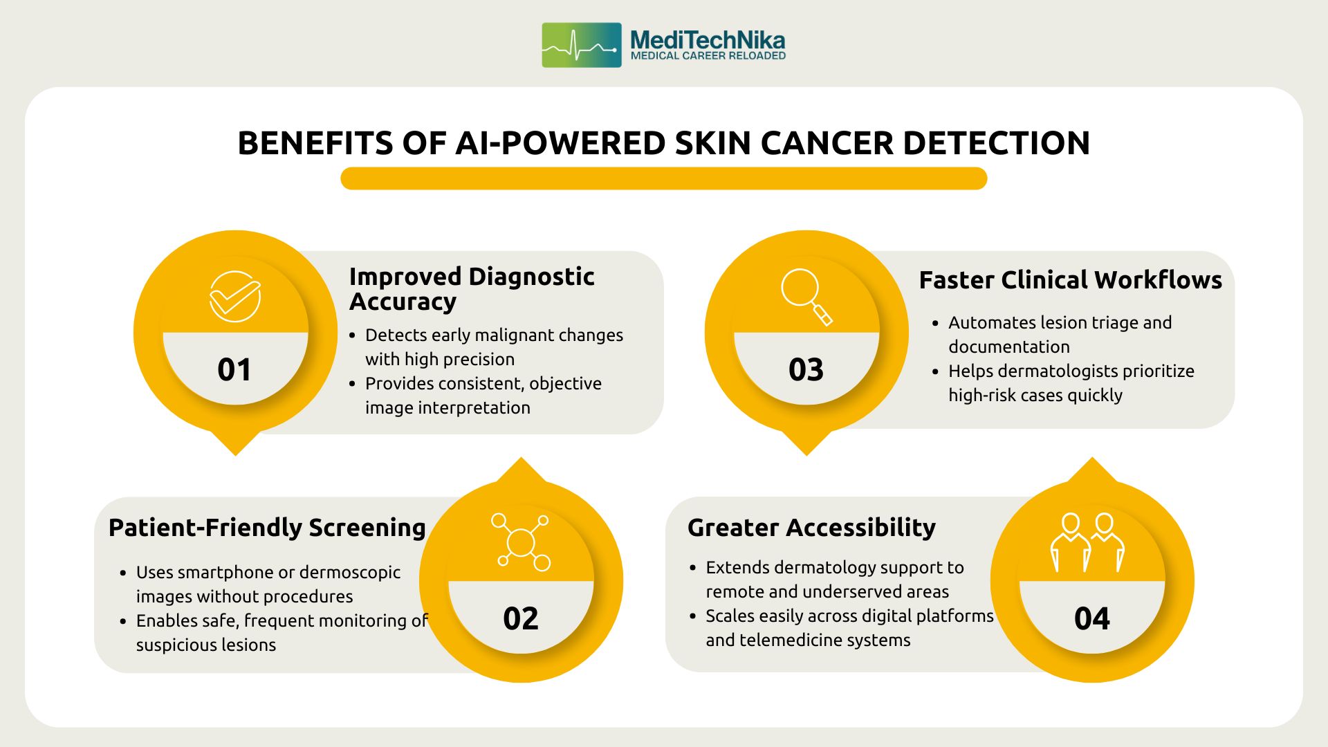
Smartphone-based AI skin analysis is not a replacement for clinicians, but it helps to broaden the reach of early detection of skin cancer to avoid further complications. It is bridging the gaps in healthcare access by integrating digital health technology.
Dermoscopy Algorithms: Precision at the Specialist Level:
Dermoscopy is also known as epiluminescence microscopy, is a technique that is used to examine skin lesions with high magnification. When this is combined with AI, dermoscopy becomes even more accurate and powerful.
Key capabilities of AI-enhanced dermoscopy:
- Automatic recognition of the structure.
- Lesion partitioning.
- Risk scoring of melanoma.
- Tracking of the skin lesions over time.
These key capabilities help dermatologists ensure accuracy and precision. AI-powered dermoscopy fits perfectly with the goals of computer-aided diagnosis, supporting clinicians but not replacing them.
Understanding dermoscopy algorithms and their principles of medical image analysis is essential for medtech students and healthcare professionals. These systems represent some of the most advanced forms of AI in healthcare.
Predictive Analytics in Dermatology: Beyond Diagnosis:
It is very crucial to identify the cancerous lesions. The next phase of digital dermatology involves prediction and prevention. With the help of predictive analytics in the healthcare sector, dermatology is creating strategic ideas for patients based on risk factors.
These systems may integrate skin type, genetics, lifestyle factors, past medical history, and UV exposure history. This helps clinicians to identify risk factors individually with the help of image-based AI and predictive modelling.
Medical Image Analysis as a Key Skill for the MedTech Generation:
For healthcare professionals and MedTech learners, mastering medical image analysis is crucial. Among other specialities like radiology, ophthalmology, and pathology, dermatology is one of the several specialities where AI is driving major transformation.
If one has to succeed in this speciality, one must master a few core skills.
- Understanding imaging standards.
- Basics of machine learning in dermatology.
- Master AI-based diagnostic tools.
- Basic understanding of data annotation and curation.
- Understanding regulatory pathways.
The future needs clinicians and engineers who can collaborate and bridge the gap between medical experience and technology.
Limitations and Challenges:
Even though there are a lot of advantages with the advanced use of AI & ML, there are a few limitations and challenges that are faced.
They are:
- Risk of algorithm bias.
- Performance differs among skin tones.
- Cannot be used without expert validation.
- Need high-quality images.
The best AI dermatology tools also need clinical verification for safe implementation.
The Future of AI and Computer-Aided Diagnosis in Dermatology:
As the technology is emerging rapidly, we can also expect a multi-modal imaging, like 3D mapping and imaging and thermal imaging. We can also expect a wearable sensor that helps to track the lesions, real-time monitoring and prediction tools, advanced dermoscopic imaging with automation, and more intelligent digital health technology.
The integration of artificial intelligence in healthcare is transforming dermatology in such a way that the transformation is from handheld smartphones to high-end dermascopes. The incorporation of AI tools in dermatology, medical imaging analysis, and computer-aided diagnosis is helping clinicians to think a way faster and also enabling more precise and accurate treatment options.
Overall, AI will constantly strengthen healthcare, delivering safer and more accurate dermatological care for patients worldwide.


